ALS community gathers at Queen’s Park to recognize the impact of the Ontario Provincial ALS Program
April 21, 2026
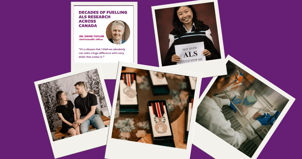
Read more
Approximately 80% of people with ALS die within two to five years of being diagnosed.


People living with ALS need help today so that they can have more tomorrows.
Approximately 80% of people with ALS die within two to five years of being diagnosed.
People living with ALS need help today so that they can have more tomorrows.
We work to improve the lives of people living with ALS by providing information and support, advocating for better care, and investing in research for a world free of ALS.
2024 Achievements
received one-on-one support from our Community Leads
engaged in 31 meetings at the federal and provincial levels
which helped fund 17 ALS research grants
Mobility wheelchairs can cost over $9,000. Our Equipment Program in Ontario changes that cost to $0.
Through our Ontario Loan Equipment Program, we provided 3,400 pieces of essential equipment in 2024, enhancing independence and quality of life for people living with ALS without emptying their bank accounts.
Join the cause and help create a better tomorrow for people diagnosed with ALS.
Fundraising events, such as Revolution Ride, plus donations and other income, contribute 84% of our funds. Without your help, we wouldn’t be half as effective at working toward a world free of ALS.

Join the cause and help create a better tomorrow for those diagnosed with ALS.
Fundraising events like Revolution Ride—plus donations and other income—contribute over 73% of our funds. Without your help, we wouldn’t be half as effective at working toward a world free of ALS.
Stay current
Join our newsletter to receive updates and stay current on what’s happening in ALS research, advocacy initiatives, support services, fundraising initiatives, events, and more.
"*" indicates required fields